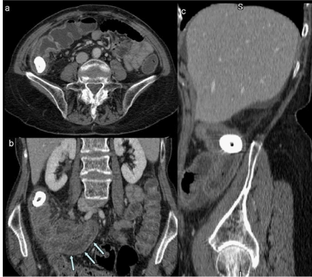
Figure 2:
Contrast enhanced CT (axial, coronal and sagittal reformats) demonstrating enterolith within a small bowel segment with proximal small bowel obstruction. Small bowel wall thickening with stratification (arrows) is also noted.