 |
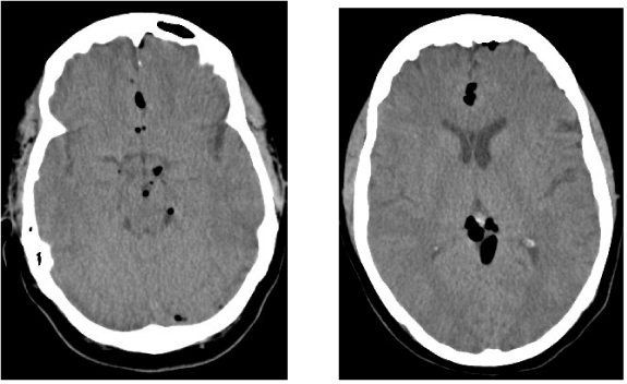
| Figure 1: Selected axial CT images of the brain done without intravenous contrast demonstrate pockets of gas within the cerebrospinal fluid of the ambient cistern (1), quadrigeminal plate cistern (2), suprasellar cistern (3), and interpeduncular cistern (4); gas is also seen within the sulci of the anterior interhemispheric fissure (5) and the left frontal region (6). These findings are compatible with subarachnoid pneumocephalus. |