|
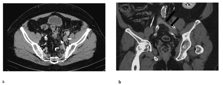
| Figure 8: a) On axial CT venogram slices, the left external iliac vein (straight white arrow) and left internal iliac vein (curved white arrow) are also dilated with enhancing walls. b) Coronal Curved MPR CT venogram demonstrates a contracted non-enhancing left common iliac segment (black arrows) leading into a dilated mid-common iliac vein. Findings are compatible with a chronic thrombosed proximal left common iliac vein with acute thrombosis of the left external and internal iliac veins. |