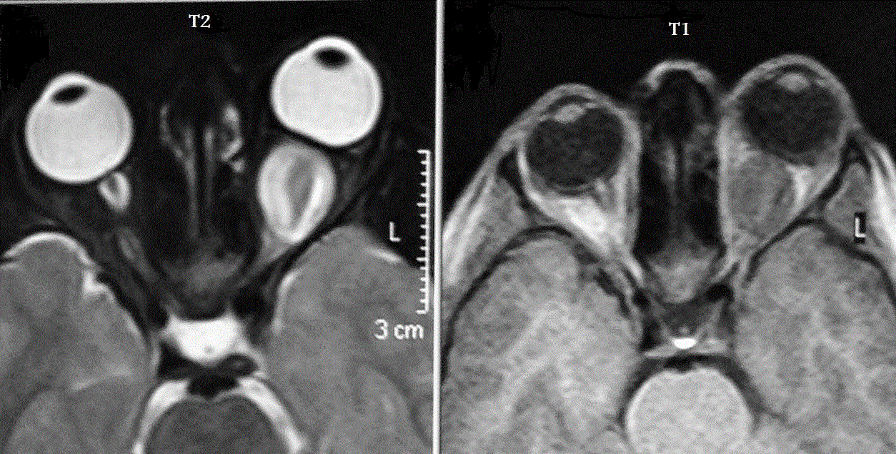
Figure 3:
T1 and T2 weighted MRI images of orbits of 8 years old girl showing left optic nerve glioma that appears hypointense on T1-weighted image and hyperintense on T2-weighted-image.