|
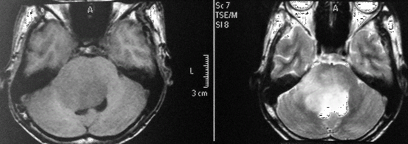
| Figure 4: T1 and T2 weighted MRI images of the brain of 22 years old male showing diffuse glioma of the pons that appears of low SI on T1 weighted image and high SI on T2 weighted and causing mass effect on the surrounding structures manifested as compression on the 4th ventricle posteriorly and encasement of the basilar artery anteriorly. |