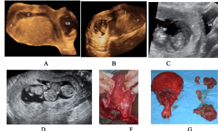
C - 3 d image showing the gestational sac with a twin pregnancy, the close localization of gestational sack to the bladder is seen.
D- Transvaginal l ultrasound showing a twin pregnancy of 11 weeks (in which the ectopic localisation is impossible to be diagnosed0.
F -Photograph of the uterine isthmus after the gestational sac has been opened surgically, which reveals the placental tissue
G- Photograph of the surgically removed uterus with the 2 foetuses
C: cavity (uterine), GS: Gestational Sac, UB: Urinary Bladder, P: Placenta, F: Fundus, T,Twin.