 |
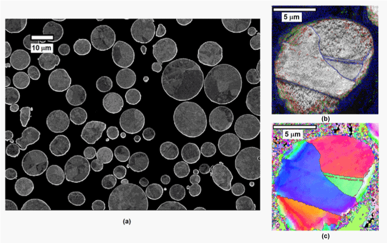
| Figure 2: SEM characterization of copper powders employed in this study. a) metallographic cross section of powders imaged with backscattered electron channeling contrast b) EBSD image of one powder particle in cross section, depicting location of high angle grain boundaries and c) Inverse pole figure map of the same powder particle. |