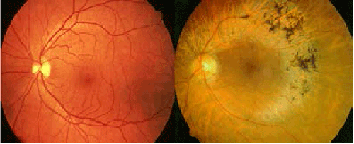
Figure 1:
Fundi of a healthy person (left) and a RP patient (right). The diseased retina exhibits peripheral intraretinal bone-spicule pigment deposit, attenuated retinal arterioles and optic disk pallor.