|
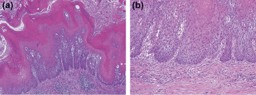
| Figure 1: Histological findings of case 1. a. The tumor show papillae with acanthosis without fibrovascular cores and tumor base is regular, broad, and pushing (magnification: ×4). b. Tumor cells are extremely well differentiated and no koilocytosis are identified (magnification: ×10). |