|
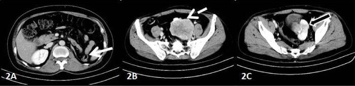
| Figure 2: CECT scan of abdomen through lumbar region shows empty left renal fossa occupied by descending colon (white arrow). Figure 2B shows a large, heterogeneous, lobulated and homogenously enhancing exophytic mass in left ectopic kidney with area of necrosis, arising in left supra and paravesical region, abutting the left lateral wall of urinary bladder ( dotted arrow). Figure 2C shows normal enhancing renal parenchyma (black arrow with white outline). |