|
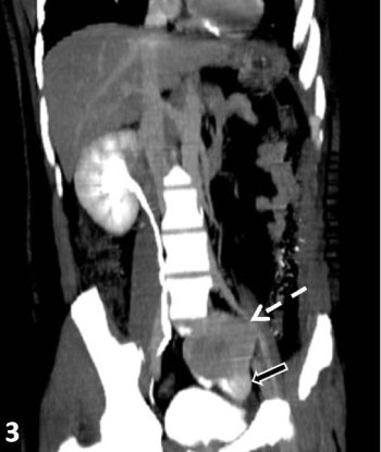
| Figure 3: CT urography image confirming the findings of axial images and shows empty left renal fossa with ectopic left kidney showing a large, heterogeneous, lobulated and heterogeneously enhancing exophytic mass with area of necrosis, arising in left supra and paravesical region, abutting the left lateral wall of urinary bladder. |