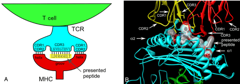
In this simplified model, CDR1 and CDR2 of TCRs mainly interact with helices running along both sides of MHC molecules, between which the presented peptide is located. In this case, CDR3 (LKVEGTRVY) interacts with a presented peptide (LDIRASELT) so as to form a ladder with 9 rungs (L–L, K–D, V–I, E–R, G–A, T–S, R–E, V–L, Y–T). For simplicity, only the interaction between CDR3 and the presented peptide is considered (SI 1). (A) AA contact energies were assigned respectively to these rungs (AA pairs) by the M-J matrix, and the binding energy between CDR3 and the presented peptide could be calculated by summing these values. (B) Actual 3-dimensional structure of a TCR–pMHC complex (PDBID: 1AO7) is shown. Both CDR3 loops comprise α and β chains running across rather than parallel to the presented peptide. CDR2 mainly interacts with helices, whereas CDR1 interacts with both MHC helix and presented peptide. The TCR–pMHC binding mode also varies widely. This image was generated using Chimera 1.5.3.