 |
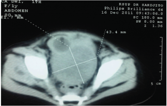
| Figure 1: CT-scan found heterogeneous mass with solid and cystic parts in uterus (oval shape, distinctive border, 41.7 × 43.3 × 82 mm3 size) that pushed urinary bladder to right anterior craniolateral and infiltrated the posterior aspect of the bladder wall. |