|
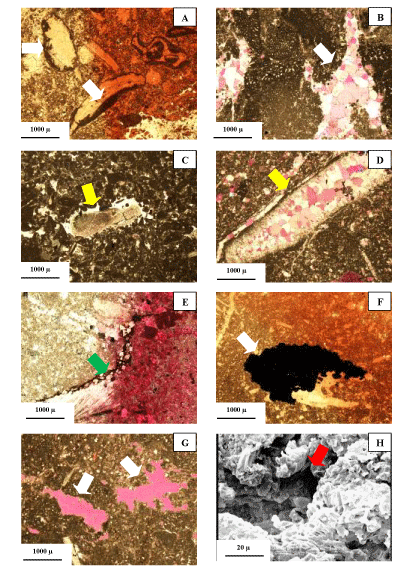
| Figure 4: Photomicrograph of different diagenetic processes. A: micrite envelope around rudist debris. PPL. B: spary calcite cement. XPL. C: syntaxial cement around echinoderm debris xpl. D: neomorphism in a skeletal debris, XPL. E: solution seams with crystals of dolomite around it. XPL. F: partial fabric selective pyritization in a shell fragment.ppl. G: vuggy porosity (in purple). XPL. H: SEM photo of microporosity. |