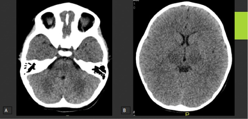
Figure 1:
1) Axial Non-Contrast CT examination of the brain at the level of the pons (A), Basal Ganglia and Thalami (B) showing subtle hypodensity along the thalami with no gross basal ganglia or posterior fossa changes.