 |
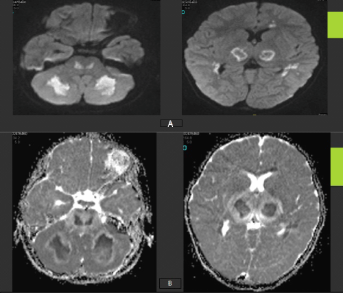
| Figure 2: Axial DWI (A) and ADC map (B) MRI brain at the level of the Pons, Basal ganglia and thalami showing bilateral rather symmetrical parenchymal areas of bright sigmal in WDI and low values in ADC maps denoting some elements of restricted diffusion which is also seen involving the corpus callosum and the cerebrellar white matter. |