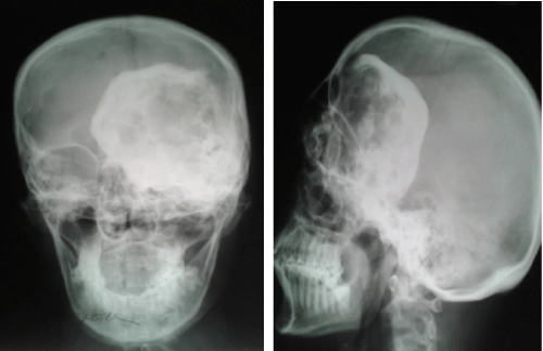
Figure 1:
Plain X ray of skull (Lateral view) in 1998, showing a fibrous dysplasia with ground glass appearance destroying the orbital cavity, frontal bone with intracranial extension.