|
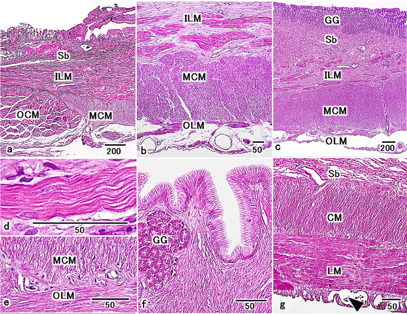
| Figure 3: LM images in the esophagus- the terminal region of the stomach. H&E stain. (a) Sagittal section of the esophagus-the cardiac region of the stomach. Mucosa, submucosa (Sb), muscularis, and serosa were observed. The muscularis was composed of an inner longitudinal (ILM), a middle circular (MCM), and an outer longitudinal layers (OLM). OCM terminated and became coincident to MCM. Scale bar=200 μm. (b) Sagittal section of muscularis in the cardiac region of the stomach. The muscularis was composed of an inner longitudinal (ILM), a middle circular (MCM), and an outer longitudinal layers (OLM). Scale bar=50 μm. (c) Sagittal section of the body region of the stomach. Mucosa, submucosa (Sb), muscularis, and serosa were observed. Gastric glands (GG) were observed in lamina-propria. The muscularis was composed of an inner longitudinal (ILM), a middle circular (MCM), and an outer longitudinal layers (OLM). Scale bar=200 μm. (d) Sagittal section of an inner longitudinal muscle layer in the body region of the stomach. Striation was observed. Scale bar=50 μm. (e) Sagittal section of a middle circular muscle layer (MCM) and an outer longitudinal muscle layer (OLM) in the body region of the stomach. Each layer was composed of smooth muscle. Scale bar=50 μm. (f) Sagittal section of mucosa in the body-terminal region of the stomach. Gastric glands (GG) in lamina-propria were disappeared from boundary. Scale bar=50 μm. (g) Sagittal section of muscularis in the terminal region of the stomach. Thin inner circular layer (CM) and thick outer longitudinal layer (LM) were observed. Subserosa (Se) was thick and the blood vessels (arrowhead) were clearly visible within them. Submucosa (Sb). Scale bar=50 μm. |