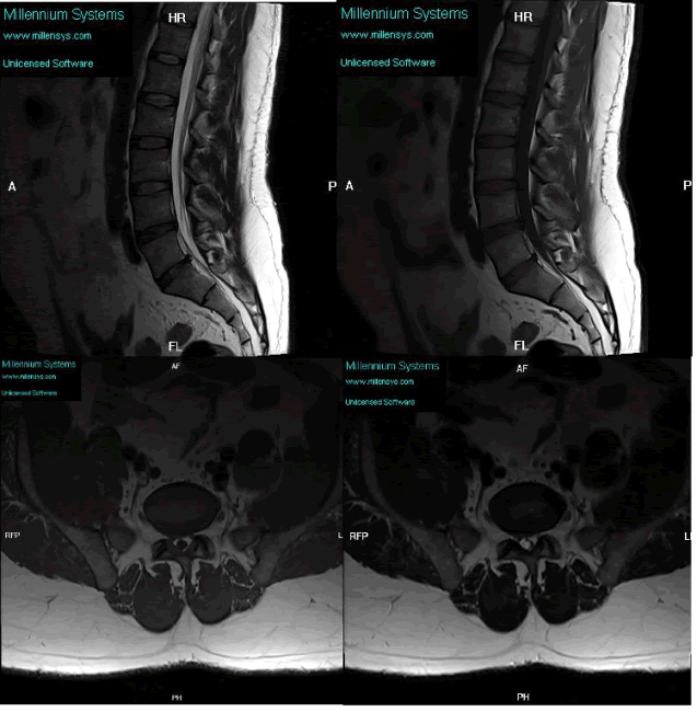
Figure 1:
T1 and T2 sagittal and axial images showed LV5-SV1 rightpostero-lateral disc protrusion compromising the right exiting andtraversing nerve roots. Also noted, LV4-LV5 mild posterior discbulge with no significant neural compromise.