|
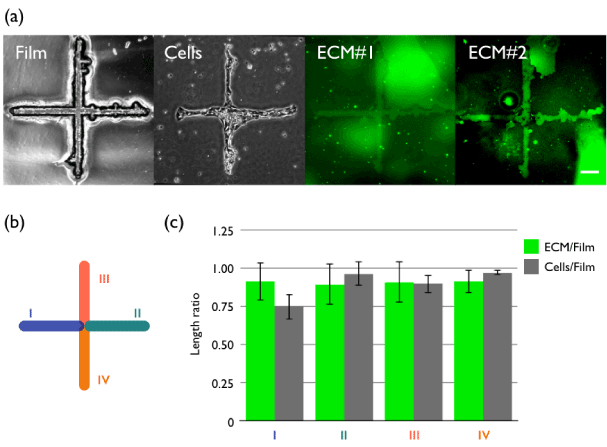
| Figure 3: Representative images of spatially selective cell adhesions by the laser-processed mask-based plasma lithography. (a) A “plus”- shaped microhole opened in an infrared absorption film (Film; phase-contrast) allows cells (Cells; phase-contrast) to selectively adhere to the unmasked regions although non-specific adhesion of fibrinogen was partly observed; here, two representative images are shown (ECM#1, #2; fluorescence). (b) Regions for evaluation of patterning accuracy. (c) The length ratios measured at the different regions (I to IV) shown in (b), represented by mean ± SD (n=3). Scale bar, 200 μm. |