 |
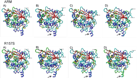
| Figure 5: Graphic representation of ARM and R157S lipases 3D structure in 10ns simulation; A) control (0ns), B) 50 °C, C) 60 °C and D) 70 °C. The figures are colored by their secondary structure, where the red indicate ß-sheets, blue represent a-helices, yellow represent 310 helices, the loops are the green and the cyan segments are random coil. The residues of Arg/Ser 157, N- and C- termini are labeled. The arrows show position of flexible regions in the structure. |