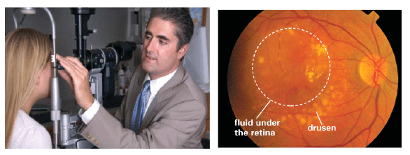
Figure 6:
Slit lamp examination (left) and retinal colour photograph shows many drusen and fluid under the retina (right) (images taken from [13]).