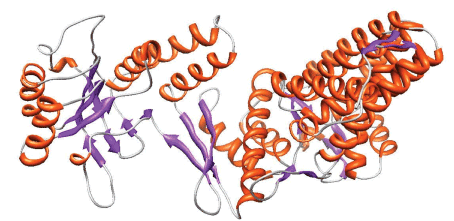
Figure 3:
The best model of the 3D structure of the Hsp60_Pb18 protein obtained by Modeller9v2 based on the alignment with 1WE3A protein. In red are the α-helices and the β-sheets are in lilac.