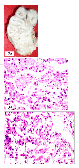
Figure 2: A: Cut section of the kidney showing all the calyces and pelvis
infiltrated by SCC. The field change pattern of pelvicalyceal involvement is
classically described for the urothelial malignancy; in SCC such involvement
may occur but is extremely rare. B: Histopathological depiction of tumor cells.
The malignant cells have completely replaced the pelvicalyceal urothelial
architecture. Few intervening vessels are seen; normal renal architecture
is obliterated. C: Microphotograph showing large malignant cells with
intervening fi brous tissue as intervening sheets. At places, squamous tumor
cells show keratinized deposition. The presence of keratin is pathognomonic
of SCC.