|
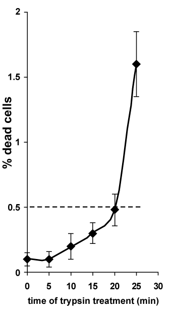
| Figure 2: HMEC viability following treatment with trypsin. In situ trypan blue staining of endothelial cell cultures at various timepoints during treatment with 0.2 µg/mL trypsin (activity 1500 U/mg). The percentage of dead cells were calculated as the average number of stained cells ± SD per field with the result from each of the five fields being averaged. The dotted line shows percent of dead cells corresponding to time which has been selected for obtaining of FCSP. |