 |
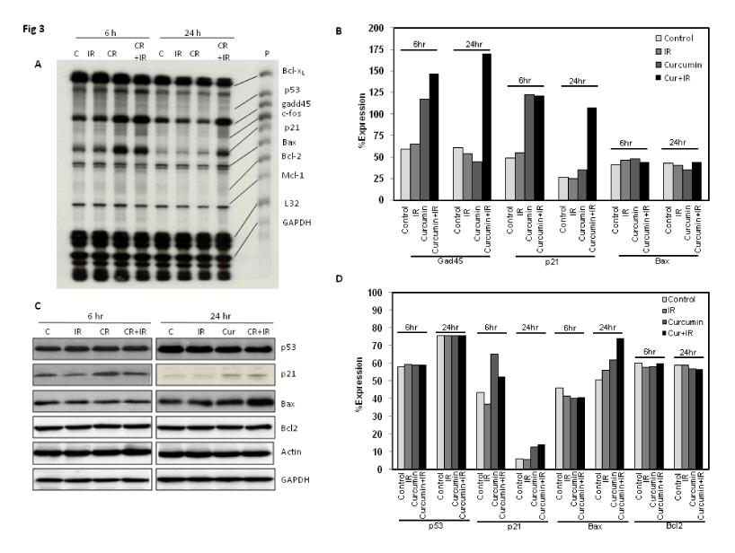
| Figure 3: Expression of p53 and p53 target genes by curcumin and radiation in PC3 cells. A: RNase protection assay showing expression of p21, gadd45, and bax at 6 and 24 hr post radiation exposure. L32 and GAPDH are loading controls. P-probe, C-control, IR- 2 Gy radiation, CR-10 ÁM curcumin, CR+IR-10 ÁM curcumin+2 Gy. B: Quantitation of RPA showing gadd45, p21, and bax expression level normalized to GAPDH. C: Western blot analysis showing expression of p53, p21, bax, bcl2 at 6 and 24 hr post radiation exposure. Actin and GAPDH are loading controls. C-control, IR- 2 Gy radiation, CR-10 ÁM curcumin, CR+IR-10 ÁM curcumin+2 Gy. D: Quantitation of Western blot showing p53, p21, bax, and bcl2 expression level normalized to GAPDH. |