|
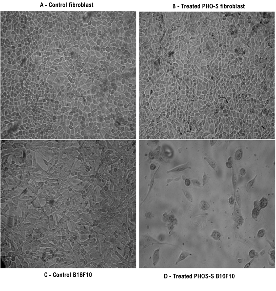
| Figure 2: Photomicrograph image obtained in inverted microscopy of control fibroblast (A) PHO-S treated fibroblast cells (B), control B16F10 melanoma cells (C) changes in the cell morphology as nuclear condensation and cell shrinkage. Similar results were obtained in three independent experiments. Image at 125X magnifications. |