|
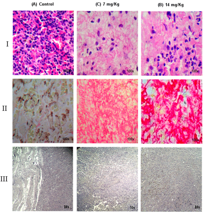
| Figure 9: PHO-S inhibition of the melanoma growth in treated mice. Representative photomicrographs of tumor sections stained with hematoxylin eosin. (I) A, B and C PHO-S decreased density of tumor cells the amount of inflammatory cells, the cellular degeneration and fibrosis. The control shows large hemorrhagic areas with numerous tumor cells. (II) A, B and C. Photomicrographs of Sirius Red-stained melanoma tumor sections from treated mice showing a large area of intratumor collagen deposition, not observed in the control. (III) A, B and C Verhoeff-van Gieson stain showed that significant decrease of elastic fibers around endothelial cells was observed in the PHO-S treated groups. |