|
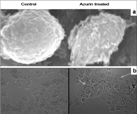
| Figure 8 :Topological analysis: (a) Scanning electron photomicrograph (SEM) of the surface of breast cancer cell ZR-75-1 treated with azurin. (b) Photomicrograph of azurin treated ZR-75-1 cells observed under light microscope at 40 X resolution. Membrane blebbing and granularity distribution of the cancer cells were shown. Arrow indicates apoptotic cells. |