|
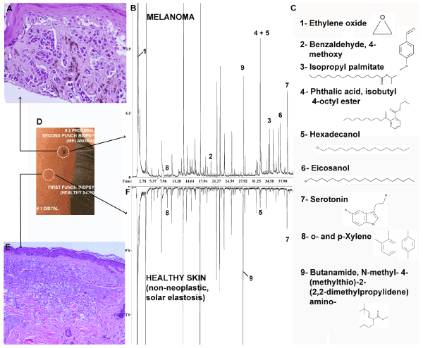
| Figure 1: Comparison of volatile signatures from a malignant melanoma biopsy and nearby healthy non-neoplastic matching skin biopsy. A. Histology - H&E staining of the #2 proximal punch biopsy melanoma lesion (40X magnification). B. Full chromatogram of melanoma sample. Some compounds found to be differentially expressed in melanoma vs skin are numbered and indicated in the chromatograms. Their names and structures are presented in C. Figure 1:. Biopsy sites from the right forearm.E. Histology - H&E staining of healthy, non-neoplastic skin showing signs of solar elastosis. F. Full chromatogram of the non-neoplastic healthy matched skin sample. |