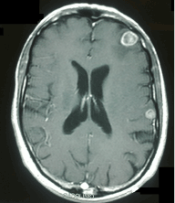
Figure 3:
Brain computed tomography showing a solid nodule of 15 mm with perilesional edema at the front left region, and a parietal superficial nodule of 7 mm.