|
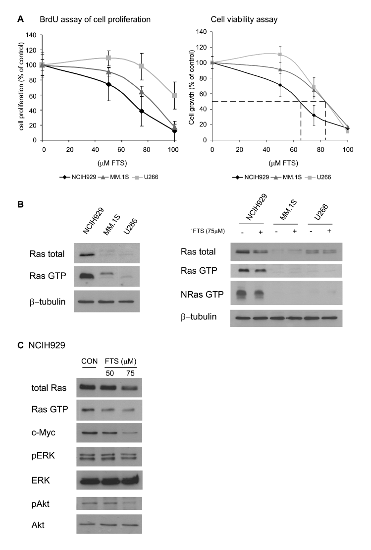
| Figure 1: Inhibition of growth of MM cell lines by the Ras inhibitor FTS. (A) Cells (2 × 10⁴ cells/well in 96-well plates) were plated and treated with 75 µM FTS or vehicle (control) for 48 h in 5% FCS medium. Cells were stained with BrdU for determination of reduction in proliferation (means ± SD, n = 3, left panel. P < 0.05 for NCIH929 and MM.1S, and P> 0.05 for U266). In another experiment, cell viability was assayed with the aid of the alamarBlue reagent (means ± SD, n = 3, right panel). (B) FTS reduces total Ras, active Ras, and NRas in NCIH929 but not in MM.1S or U266 MM cell lines. Cells (1 × 10⁶ cells/ml) were grown in 5% FCS medium for 72 h and then lysed (left panel). Alternatively, cells (1 × 10⁶ cells/ml) were plated in 5% FCS medium and treated with 75 µM FTS or vehicle (control) for 72 h and then lysed (right panel). Ras activity was determined by a GTP-Ras pull-down assay followed by western blotting with anti-pan-Ras or anti-NRas antibodies. The amounts of Ras and ß-tubulin were quantitatively analyzed by western immunoblotting. Typical immunoblots visualized by ECL are shown (n = 3). (C) Ras downstream effectors are inhibited by FTS in NCIH929 cells. NCIH929 cells were grown and treated as described in B. Active Ras, total Ras, c-Myc, p-ERK, ERK, p-Akt, and Akt were quantitatively analyzed by western immunoblotting. FTS at 75 µM reduced the amount of proteins by 56 ± 4% for Ras-GTP, 41.8 ± 7% for total Ras, 65 ± 2% for c-Myc, 38 ± 5% for pERK, and 52 ± 1.6% for pAkt in (means ± SD, n = 3). |