 |
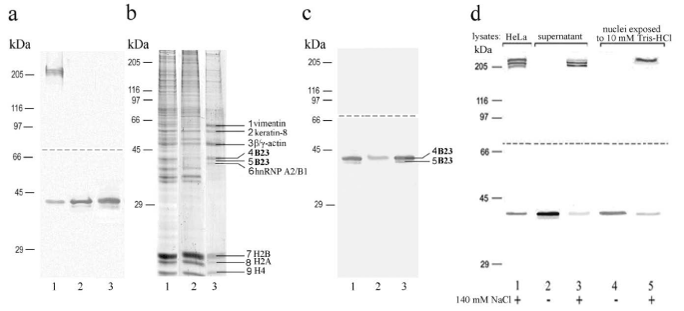
| Figure 2: Immunochemical (a, c, d) and electrophoretic (b) analysis of the influence of hypotonic treatment of HeLa cells (a) and nuclei (c, d) on state of nucleophosmin. a) HeLa cells in PBS (1), RSB (2), nuclei in RSB (3); b, c) nuclei in RSB (1), fraction of nuclei (2) and supernatant (3) after extraction with 10 mM Tris-HCl; d) HeLa cells (1), fractions of supernatant and nuclei were kept in 10 mM Tris-HCl (2, 4), or in 10 mM Tris-HCl, containing 140 mM NaCl (3, 5). Samples were treated for electrophoresis in buffers (PBS, RSB, or 10 mM Tris-HCl) containing 30% glycerol by lysing solution at 100°C for 1 min. SDS-PAGE was performed by the Laemmli method in 7.5% PAG. The proteins were electrotransferred in two stages. Positions of the nine protein bands of supernatant fraction more intensively stained with Coomassie G-250 are shown on the right (b); the two of them (4 and 5) contained nucleophosmin (c, lane 3). |