 |
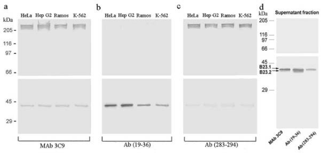
| Figure 4: Immunochemical analysis of specificity of MAb 3C9 (a), Ab (19-36) (b) and Ab (283-294) (c). Tumor cells (a-c) and supernatant fraction obtained after nuclei treatment (d) were used. SDS-PAGE was performed by the Laemmli methods in 7.5% (a-c) and 10% PAG (d). Samples were treated as described on legend to Figure 1b. |