|
|---|
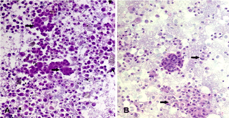
| Figure 5: A) Smears prepared from HepG2 cells showing malignant hepatocytes forming acini and clusters of cells with enlarged nuclei and increased nucleocytoplasmic ratio, arrows (H&E, 200). B) HepG2 cells after treatment with Terminalia bellerica methanol extract showing small shrinking hepatocytes with apoptotic and degenerative changes, arrows (H&E, 200). |