 |
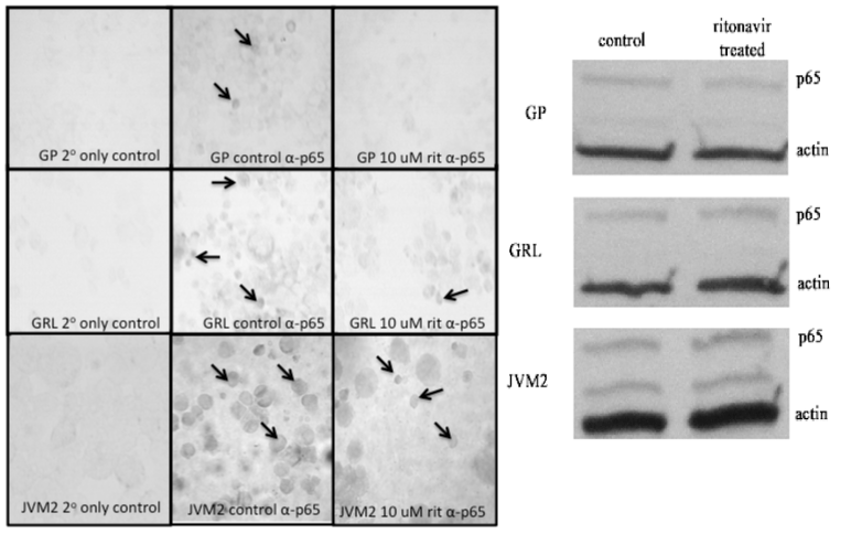
| Figure 3: Effects of ritonavir treatment NFκB protein levels in MCL. Effects of 24 hours treatment with 10 μM ritonavir on the protein level expression and localization of NFκB subunit p65 in GP, GRL, and JVM2 cell lines, as assessed using immunocytochemistry (left panel, 40X magnification) and western blotting (right panel). GP, GRL, and JVM2 cells (1 x 10⁶ cells) were treated for 24 hours with 10 μM ritonavir. For immunocytochemistry, cytospins were prepared and staining performed for p65. For western blotting, protein was then isolated with cell lysis buffer, run on SDS-PAGE gels, transferred to PVDF membranes, and blotted for p65. Arrows on immunohistochemistry figure indicate representative nuclear staining of p65. |