|
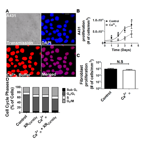
| Figure 3: Association therapy affects cell cycle progression. A. Representative image of adenovirus infection on A431 cells. Superior panels show transmission image of A431 cells and nuclei stained with DAPI (blue). Left inferior panel shows expression of the Ca2+ buffering cassette (red). Merged image on right inferior panel confirms expression of nuclear Ca2+ buffering in 100% of the cells. B. Cell growth curve of A431 cells 24, 48, 72 and 96 h in control conditions and after infection with a type V adenovirus construct that buffers Ca2+n signaling. Proliferation is decreased upon Ca2+n buffering compared to control, (p<0.0001, t test was used for each day of experiment, and one-way analysis of variance, Bartlett’s test for equal variances, for all groups, N = 3). C. Cell proliferation of human gingival fibroblast at 96 h under control or Ca2+n buffering condition. There were no significant changes in cell proliferation between control and fibroblasts that had Ca2+n buffered (N = 3). D. Cell cycle phase analysis of A431 cells under control conditions, XRCd10Gy, Ca2+n, and after Ca2+n + XRCd10Gy conditions. Under association therapy the number of cells in G2/M phase increased compared to control. The data are expressed as mean ± SEM of triplicate measurements and are representative of N = 3 experiments (p<0.05, one-way analysis of variance). |