|
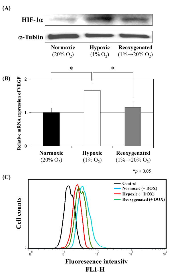
| Figure 2: Effect of altering oxygen conditions on human MFH cells in vitro. A human MFH cell line, Nara-H was incubated for 48 h under one of three different oxygen conditions: normoxic (20% O2, 5% CO2, 75% N2); hypoxic (1% O2, 5% CO2, 94% N2); or reoxygenated conditions. In the reoxygenated condition, cells were incubated under normoxic conditions for 24 h followed by 24 h exposure of hypoxic conditions. (A) Immunoblot analyses showed that HIF-1a expression was increased in hypoxic conditions compared with normoxic conditions, but was decreased in reoxygenated conditions. (B) mRNA expression of VEGF in cells cultured in one of three different oxygen conditions was evaluated by qRT-PCR. VEGF mRNA expression was significantly increased in hypoxic MFH cells compared with normoxic and reoxygenated cells (*p < 0.05 versus both normoxic and reoxygenated conditions). (C) Apoptotic effect of DOX treatment on human MFH cells under three different oxygen conditions was assessed DNA fragmentation assay by FACS analyses. Under normoxic conditions, DOX treatment induced apoptosis of human MFH cells (light blue). Apoptotic activity was strongly decreased in hypoxic cells (red), and the activity was recovered under reoxygenated conditions (green). |