|
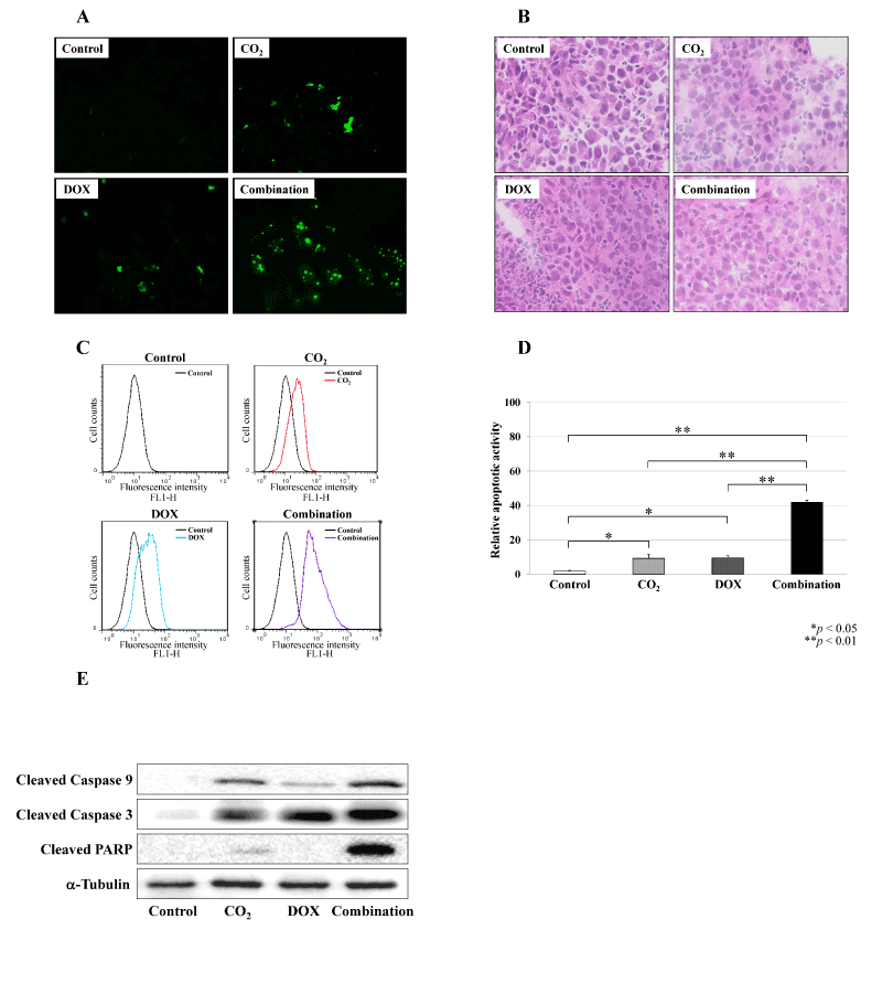
| Figure 5: Effect of the combination therapy using CO2 treatment and DOX on human MFH cell apoptosis. We examined the apoptotic activity in treated tumors by immunofluorescence staining, DNA fragmentation assay and immunoblot analysis. (A) Immunofluorescence staining showed that more apoptotic cells were detected in the CO2 or DOX groups compared with the control group, whereas the number of apoptotic cells was dramatically increased in the combination group. (B) H&E staining matching the immunofluorescence staining. (C) DNA fragmentation was assessed by flow cytometry in the control (black), CO2 (red), DOX (light blue), and combination groups (purple) at the end of treatment (14 d). (D) Fluorescence intensity in each group was normalized to that in control group, and statistically analyzed. Consistent with the immunofluorescence results, apoptotic activity in the CO2 and DOX groups was significantly increased compared with that in control group (*p < 0.05). The apoptotic activity in the combination group was significantly increased compared with the control, CO2 and DOX groups (**p < 0.01). (E) Immunoblot analyses determined that increased expression of the cleavage products of caspase 3, 9 and PARP occurred in the CO2, DOX, and especially the combination group compared with the control group. |