 |
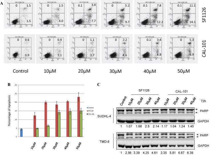
| Figure 2: Apoptosis induction by SF1126 and CAL-101 inB-NHL cell lines. (A). SUDHL-4 cells were treated with SF1126 or CAL-101 at different doses [10 然, 20 然, 30 然, 40 然 and 50 然] for 72-hr. Apoptosis was detected by flow cytometry based on propidium iodide (Y-axis) and annexin V staining (X-axis). Percentages of apoptotic cells (lower right quadrant represents the early apoptotic population, upper right quadrant represents the late apoptotic population and upper left quadrant represents the necrotic population) are indicated. (B). TMD-8 cells were treated as above and apoptosis was analyzed by flow cytometry after staining of propidium iodide and annexin V. The graph represents the mean percentage of apoptosis S.D. (n = 3). (C). SUDHL-4 and TMD-8 cells were treated with SF1126 and CAL-101 at 10 然, 20 然, 30 然 and 40 然 for 72 hr. Apoptosis was evaluated by immunoblotting to detect PARP cleavage with an anti-PARP antibody. GAPDH was used as a loading control. Numbers indicates density of cleaved PARP. |