|
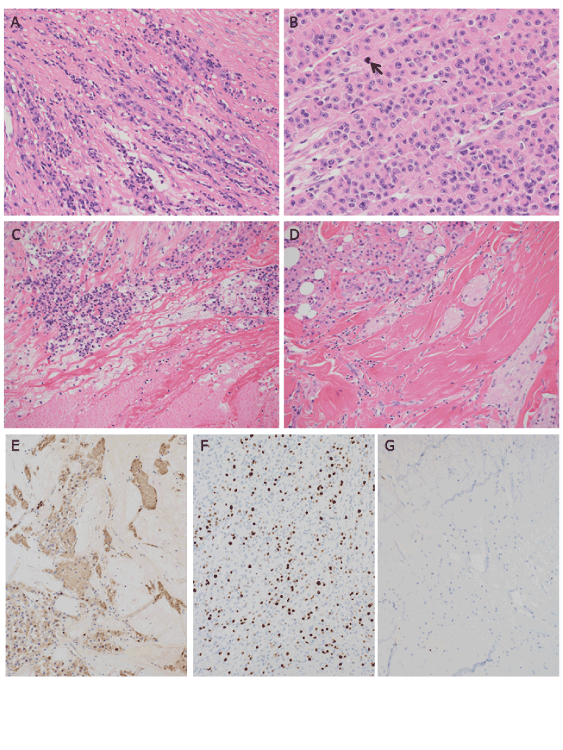
| Figure 1: Histological findings of the malignant granular cell tumor A) The tumor cells were proliferating in a fascicular or trabecular fashion (H.E. staining: original magnification x 100). B) The tumor cells were tightly packed, and the nuclear-to-cytoplasmic ratio was high. The tumor cells had eosinophilic granular cytoplasm with vesicular and prominent nucleoli. Nuclear pleomorphism was evident. Mitoses were also seen (arrow) (H.E. staining: original magnification x 400). C) Tumor necrosis was also noted (lower part) (H.E. staining: original magnification x 100). D) Small areas of benign granular cell tumor were observed at the periphery of the tumor (lower right), adjacent to the extensive malignant area (H.E. staining: original magnification x 100). E) Immunohistochemical expression of S-100 protein was observed at the borderline area between benign (upper right) and malignant (lower left) areas (Immunohistochemistry: original magnification x 100). F) Ki-67 index in the malignant area was up to 40% in the highly malignant area (Immunohistochemistry: original magnification x 100). Ki-67 positive cell could be seldom seen in the benign area at the periphery (Immunohistochemistry: original magnification x 100). |