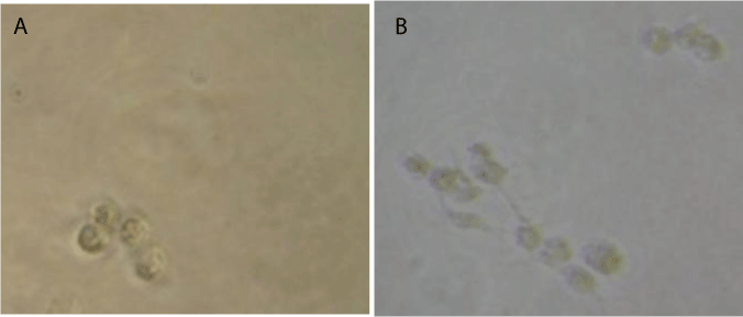
Figure 2:
MCF-7 cells exposure to (A) FeCp
2
3000 μM or (B) FeCp
2
BF
4
300 μM during 24h. Cells were visualized by inverted microscope. Magnification, 10X, insets, 40X.