 |
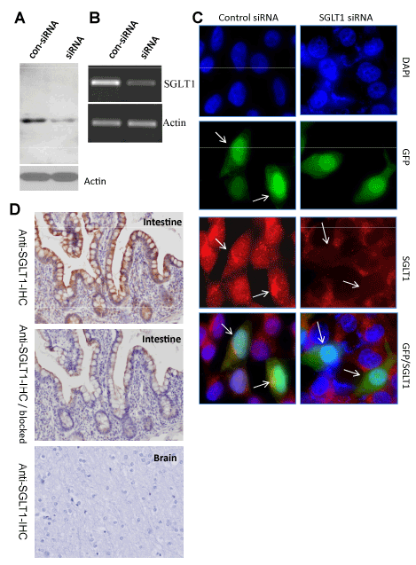
| Figure 2: Characterization of the SGLT1-IHC antibody using HCT116 human colon cancer cells. A, Western blot analysis of endogenous SGLT1 in cells transfected with control shRNA or SGLT1 shRNA using the SGLT1-WB antibody, which was consistent with the changes of SGLT1 mRNA levels determined by RT-PCR B, Actin was used as internal controls. C, Immunocytochemical analysis of cells transfected with control shRNA or SGLT1 shRNA using SGLT1-IHC. Cells containing shRNA appear green (arrows) due to the expression of GPF under the control of an autonomous promoter and the SGLT1 signal is red. Nuclei are stained blue with DAPI. D, Immunohistochemical analysis of a human normal intestine sample using SGLT1-IHC with or without blocking peptides (1 mg/mL). |