|
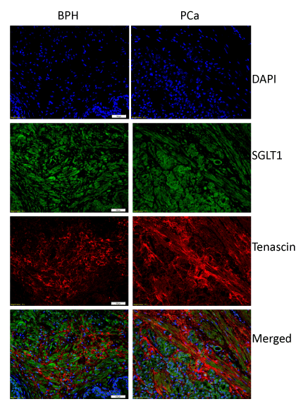
| Figure 6: SGLT1-positive stromal cells are negative for tenascin. Immunofluorescent co-staining of SGLT1 (green) and tenascin (red) on tissue samples of BPH (left) and PCa (right). Nuclei were stained by DAPI (blue). Co-expression of SGLT1 and tenascin would have appeared yellow. Bar=50 μm. |