| Figure 3: [A-H]: Morphological features of apoptotic MCF-7 cells post treatment with 50% inhibitory concentration (IC50) of both C.cerastes and V.lebetina venoms compared to untreated cell control using transmission electron microscope (TEM). [A and E]: Untreated cell control; [B, C, D]: cells treated with C.cerastes (1.5 μg/ml); [F, G, H]: cells treated with V.lebetina (2.5 μg/ml). Treated cells exhibited characteristic features of apoptosis as chromatin condensation, cytoplasmic vacuoles, cell membrane budding as well as undefined mitochondrial cristae structure. |
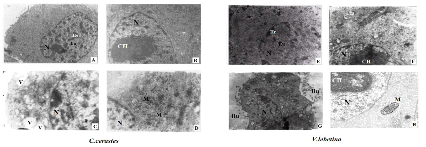 N: Nucleus; He: Heterochromatin; Eu: Euchromatin; M: Mitochondria; CH: Chromatin condensation; V and vertical arrows: Vacuoles; Bu: Cell membrane budding.