|
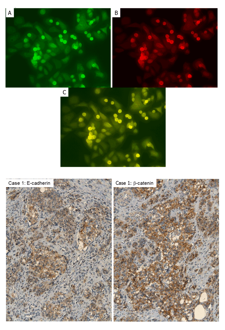
| Figure 3: Immunofluorescence using HeLa cells. A) Mergedphotograph (C) shows that FGFR4 expression (A: red) is restricted to EWS-WT1(-KTS) transfected cells (B: green). B) Immunohistochemical analysis of DSRCT clinical sample. E-cadherin and β-catenin membranous expression was seen in DSRCT case (case 1). Note that some cytoplasmic staining of β-catenin is also seen. |