 * Indicates significant p value.
|
* Indicates significant p value. |
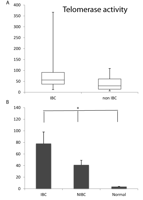
| Figure 3: (A) Box plot of Telomerase activity values in IBC and non IBC. Minimum and maximum values of telomerase activity are depicted by upper and lower bars, the box signifies the upper and lower quartiles, and the median is represented by a short black line within the box. (B) Histogram of the mean telomerase activity in IBC, non IBC and normal tissue samples where there is a significant difference (P<0.001). |