 |
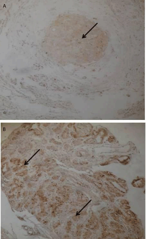
| Figure 6: Immunohistochemical staining using anti-hTERT sera of a (A) non inflammatory and (B) of inflammatory breast tissue sample. hTERT positive tumor cells showed brown nuclei (arrows) by 3,3’-diaminobenzidine (DAB) according to the protocol described previously Original magnifications, X100. |