|
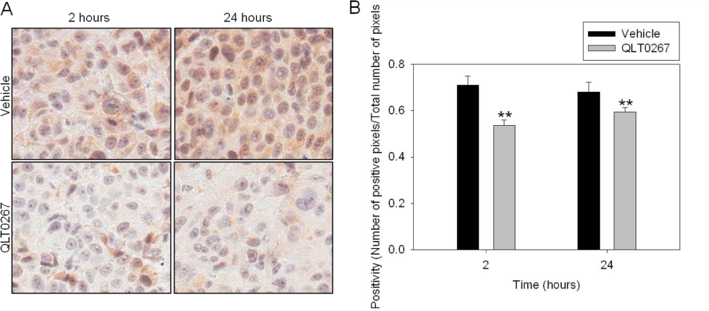
| Figure 4: Kinetics of p(ser473)AKT expression in vivo. Representative p(ser473)AKT IHC images from tumors collected after 2 and 24 hours of vehicle and QLT0267 treatment are shown (A). Quantification of p(ser473)AKT positive pixels in TMA tumor cores from vehicle and QLT0267 treated animals was performed using the Aperio Image Scope software. A decrease in positivity of p(ser473)AKT in the QLT0267 treated tumors is seen when compared to the vehicle control tumors at 2 (P<0.005) and 24 hours (P<0.005) (B). |