|
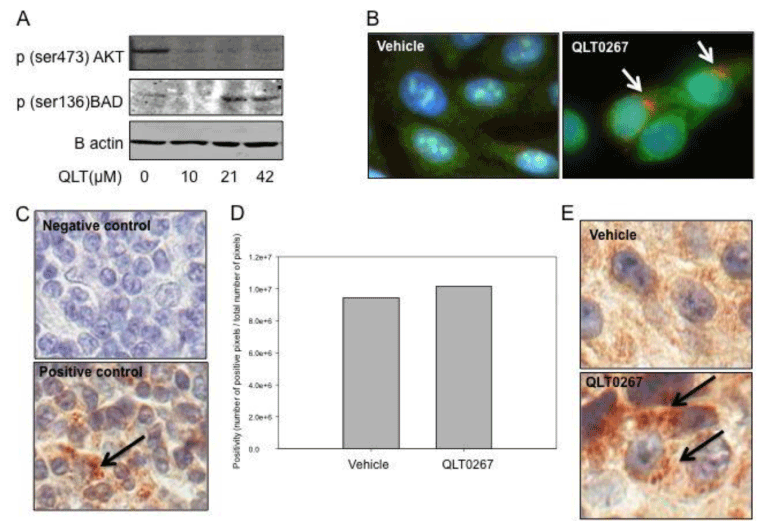
| Figure 5: QLT0267 triggers a change in subcellular localization of the pro-apoptotic protein BAD. In vitro examination of p(ser136)BAD expression shows that QLT0267 treated LCC6WT-luccells have decreased expression of p(ser473)AKT and increased expression of activated BAD p(ser136)BAD (A). Immunofluorescence of BAD (red) and Bcl-xl (green) was examined after treatment. An alteration in the subcellular localization of BAD is observed (white arrows) (B). In vivo, the negative and positive control staining for BAD is provided using tonsil tissue (C). Primary antibody was omitted in the negative control. The positive control shows that BAD are localized to punctate structures in the cytoplasm (black arrow). BAD expression was assessed in tumors from animals treated with vehicle or QLT0267. Staining was quantified using the Aperio Image Scope software (D). A small increase in BAD expression was observed in QLT0267 treated tumors (D). Representative tumor cores from the 2 hour time-point for vehicle treated (E) and QLT0267 treated (E) tumors, are shown. In the tumors from animals treated with the vehicle control BAD distribution appears to be diffuse and cytoplasmic whereas in tumor tissue from animals treated with QLT0267 BAD appears to be localized to puncta (black arrows). |